- Misión
- Equipo de Trabajo
- Servicios
- Galería
-
Libro Tópicos.
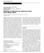
Fotos de L3A de Gnathostoma
|
|
|
|
||
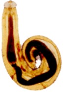
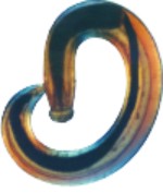
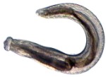
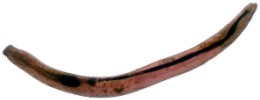
Fig.1. Larva L3A recuperada del ojo de una paciente.
Fig.2. Larva L3A obtenida del músculo esquelético deEgretta alba.
Fig.3. Larva L3A aislada de la masa muscular deDormitator latifrons.
Fig.4. Larva L3A recuperada de una biopsia excisional de un paciente.
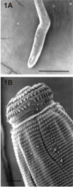
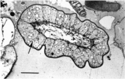
Fig. 1A, 1B Scanning electron micrographs of AL3 larvae recovered from the liver of a cat infected with 20 AL3 larvae from Egretta alba. A Whole larva. Bar=1 mm. B Head bulb and upper body, with papilla (arrowhead) between rows 12 and 13. Bar=100 um.
Fig. 2 Light micrograph of a 1 lm section through the intestine of an AL3 larva from Pelecanus occidentalisstained with toluidine blue. Intestinal cell nucleus (arrow). Bar=30 um.
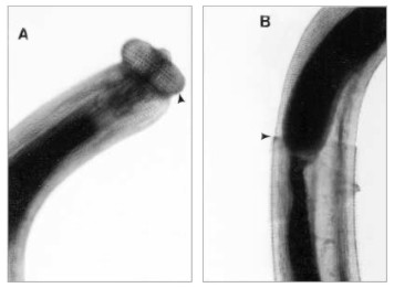
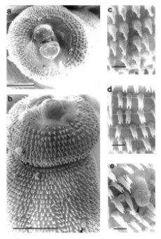
Fig. 4A Light micrograph of a whole young adult worm recovered from thoracic muscle of experimental cat. A Nine rows of hooklets on the head bulb (arrowhead) and B the rim of the cuticle (arrowhead) midway on the body. Bar=500 um.
Fig. 5A-E Scanning electron micrograph of a young adult worm from the stomach of an experimentalcat. A View showing the head bulb with an incomplete first row of hooklets. Bar=100 lm. BHead bulb with nine rows of single-pointed hooklets and body spines. Bar=100 lm. CHigher magnification of single-pointed body spines in first two rows. D Bifurcated spines, E trifurcated cuticular spines with cervical papilla between rows 15 and 16. Bars in C-E= 10 um.
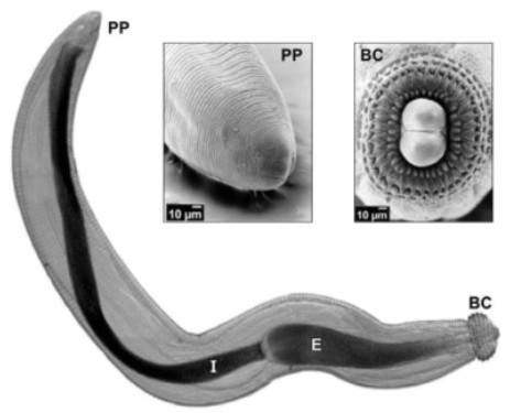
Fig. 31.1. Larva L3A deGnathostoma spprecuperada de carne de pescado y observada mediante microscopía de luz y electrónica de barrido: BC, bulbo cefálico; E, esófago; I, intestino y PP, parte posterior del cuerpo. (Las fotomicrografías de barrido fueron tomadas por José Guadalupe Rendón Maldonado, CINVESTAV-IPN).
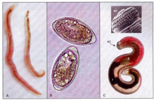
Fig. 47-1 A, Adultos hembra y macho de Gnathostoma sp, recuperados de una cavidad parasitaria del estómago de Didelphis virginiana (tlacuache). B, Huevos de Gnathostoma sp. miden 65 x 40 um y presentan un cuerpo polar más prominente. C, Larva L3A de Gnathostoma sp. Obtenida de Dormitator latifrons, 3 a 4 mm de longitud. BC Bulbo cefálico con hileras concéntricas de ganchos. E, esófago; I, intestino.
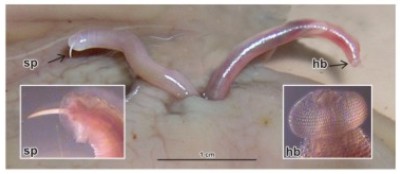
Fig. 7.1
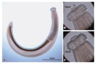
Fig. 7.2
Figure 7.1. Adult worms ofGnathostoma in stomach wall of opossum (Didelphis virginiana) with enlarged spicule (sp) (left) and head-bulb (hb) (right). See also color insert.
Figure 7.2. Advanced third-stage larva of Gnathostoma spinigerum. (A) Whole worm. (B) Anterior part showing lips and papillae (arrow). (C) Anterior part showing hooklets and body spines. See also color insert.
PUBLICACIONES
|
|
|
|
|